The University of British Columbia boasts a wide range of advanced imaging equipment available to researchers across various disciplines. This webpage provides an overview of the available technologies and links to the respective facilities that house them.
In Vitro Techniques
- Confocal Microscopy: Laser scanning confocal microscopy delivers high-resolution 3D cellular and tissue imaging
- Correlative Light and Electron Microscopy (CLEM): Combine light and electron microscopy for a comprehensive view of your samples.
- Conventional Optical Microscopy: Utilize standard light microscopes for a variety of biological applications.
- Cryo-Electron Tomography (Cryo-ET): Image frozen, hydrated samples in 3D at near-molecular resolution.
- Cryo-Transmission Electron Microscopy (Cryo-TEM): Image frozen, hydrated samples in 2D at near-molecular resolution.
- Electron Probe Microanalysis (Electron Microprobe): Analyze the elemental composition of your samples.
- Electron Tomography: Generate 3D reconstructions of your samples using transmission electron microscopy.
- Focused Ion Beam Scanning Electron Microscopy (FIB-SEM): Combine high-resolution imaging with the ability to mill and manipulate your sample at the nanoscale.
- Multi-Photon Microscopy: Image deep tissues with minimal photobleaching.
- Nuclear Magnetic Resonance Spectroscopy (NMR): Atomic-level insights into molecular structures and dynamics.
- Particle Size Analysis (Mastersizer): Precisely characterize particle size (nm-mm).
- Scanning Electron Microscopy with Energy-Dispersive X-ray Spectroscopy (SEM/EDX): Image your samples and analyze their elemental composition.
- Super-Resolution Microscopy: Achieve resolutions beyond the diffraction limit of conventional light microscopy.
- Transmission Electron Microscopy (TEM): Image your samples at high magnifications for detailed structural analysis.
- Ultra-High Performance Liquid Chromatography-Mass Spectrometry (UHPLC-MS/MS): Unparalleled sensitivity for accurate trace compound quantification.
- X-ray Diffraction: Determine the crystal structure of your samples.
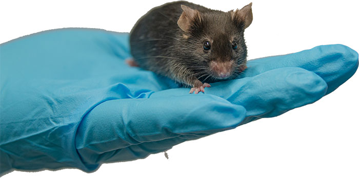
In Vivo Techniques
- In-Vivo Imaging System (IVIS): Visualize in vivo processes via high-sensitivity bioluminescence/fluorescence.
- MR Imaging: Achieve high-resolution anatomical, microstructural, and functional imaging with the 9.4T preclinical MRI scanner.
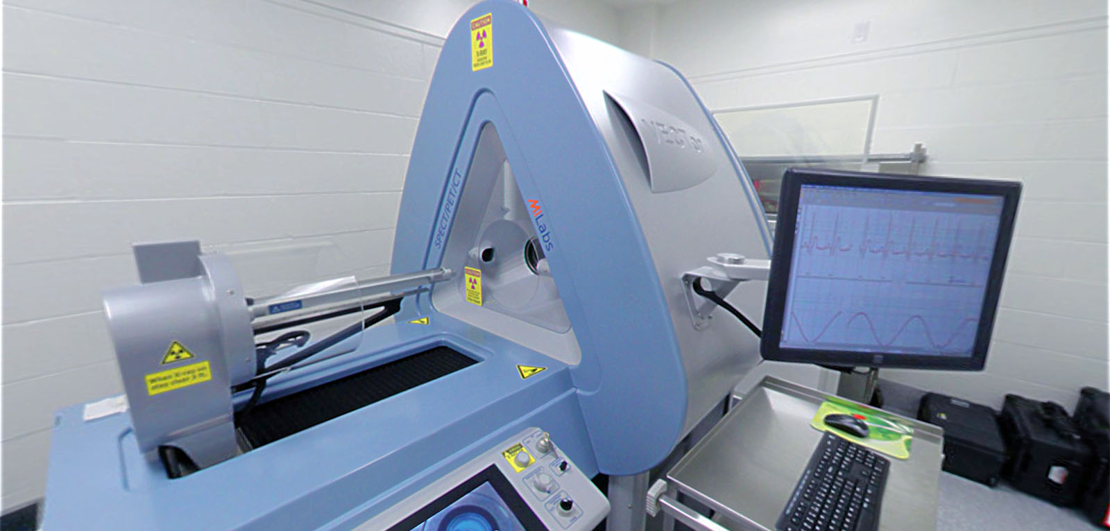
- PET/SPECT/CT: Utilize Positron Emission Tomography (PET), Single-Photon Emission Computed Tomography (SPECT), and X-ray Computed Tomography (CT) for advanced molecular imaging.
- X-ray Imaging: Utilize X-rays for non-destructive imaging of your samples.

Sequencing & Genotyping
- Genotyping Facility, School of Biomedical Engineering: Access state-of-the-art equipment and expertise for your genotyping needs.